Red Blood Cell Physiology, Hemoglobin Production, Hemoglobin A2, Hemoglobin A, Hemoglobin F, Hemoglobin H, Hemoglobin Bart
- Physiology
- Erythropoietin
- Primary Hormone regulator of RBC production
-
Erythropoietin sources
- Fetus: Monocyte and Macrophage system in liver
- Postnatal: Peritubular cells in Kidney
- Physiology
- Red Blood Cell
- Hemoglobin produced until amounts to 90% of RBC mass
-
Red Blood Cells start as Reticulocytes in Bone Marrow
- Reticulocytes are juvenile Red Blood Cells
- Nucleus extruded once RBC has matured
- Reticulocytes contain ribosome remnants
- Immature Reticulocytes contain most ribosomes
- Mature Reticulocytes contain least ribosomes
- Reticulocytes have 4 day life span
- Bone Marrow: 3 days (less if Erythropoietin high)
- Peripheral blood: 1 day
- Reticulocytes are juvenile Red Blood Cells
-
Red Blood Cell survival
- Normal RBC: 120 days
- Abnormal RBC: May survive as little as 15 days
- Following transfusion: RBC survival 2-3 weeks
- Physiology
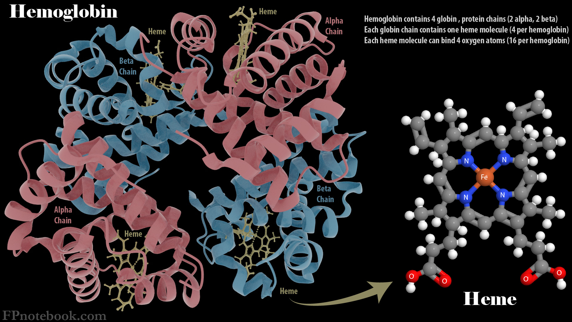
- Hemoglobin
- Normal Hemoglobin is composed of 4 Protein-Heme complexes
- Two pairs of polypeptides (4 total)
- A pair of alpha chains are found in every normal Hemoglobin type
- A pair of other identical polypetides depending on Hemoglobin type (Gamma, Beta, Delta)
- Central iron-containing heme ring
- Attached to each of the 4 polypeptides
- Images
- Two pairs of polypeptides (4 total)
- Six types of normal Hemoglobin
- Embryonic
- Gower I
- Gower II
- Portland
- Fetal Hemoglobin (HbF): Alpha2-Gamma2
- Primary Hemoglobin in fetus
- Replaced by Adult Hemoglobin by age 6-12 months
- Adult Hemoglobin (HbA): Alpha2-Beta2
- Adult Hemoglobin (HbA2): Alpha2-Delta2
-
Thalassemia related Hemoglobins
- Hemoglobin Bart's: Gamma4
- Seen in Alpha Thalassemia
- Hemoglobin H: Beta4
- Seen in Beta Thalassemia
- Hemoglobin Bart's: Gamma4
- Sickle Cell Related Hemoglobin (Hb S)
- Hemoglobin S (Hb S) replaces the normal Hemoglobin A
- Deoxygenated Hemoglobin-S assumes a sickle shape deforming Red Blood Cells
- Deoxygenated HbS aggregates under low oxygen tension
- Molecules polymerize into a gelatinous network
- Deforms Red Blood Cells into a sickle shape
- Red cells with sickle shape are less deformable
- Chromosome 11 Mutation: Substitution of Amino AcidValine for Glutamic Acid
- Occurs at the 6th position of the Hemoglobin beta-chain
- Results in a "sticky" Hemoglobin that forms a rigid chain when deoxygenated
- Sickle cell gene is inherited in Autosomal Recessive pattern
- Heterozygotes have Sickle Cell Trait and Homozygotes have Sickle Cell Anemia
- Sickle Cell Trait is protective against Malaria, resulting in up to 40% trait Prevalence in some African regions
- Hemoglobin S (Hb S) replaces the normal Hemoglobin A
-
Hemoglobin Metabolism
- Hemoglobin breakdown occurs when Red Blood Cells are destroyed (e.g. in Spleen at the end of their roughly 120 day life cycle)
- Hemoglobin is converted into Biliverdin and ultimately into Bilirubin for excretion
- See Bilirubin
- Resources
- MMWR Iron Deficiency Anemia Prevention