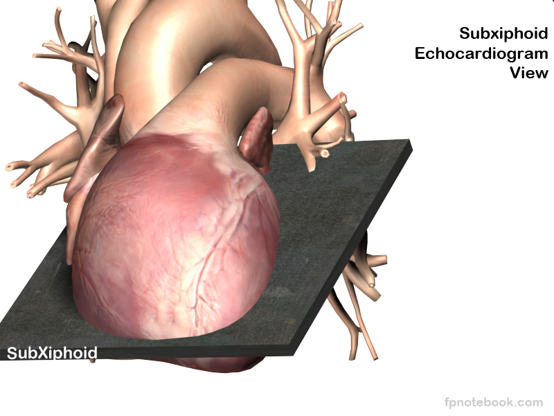
Subcostal Echocardiogram View, Subxiphoid Echocardiogram View
- See Also
- Echocardiogram
- FAST Exam
- Parasternal Long-Axis Echocardiogram View ( PLAX View)
- Parasternal Short-Axis Echocardiogram View (PSAX View)
- Subcostal Echocardiogram View (Subxiphoid Echocardiogram View)
- Apical Four Chamber Echocardiogram View
- Suprasternal Echocardiogram View
- Echocardiogram in Congestive Heart Failure
- Inferior Vena Cava Ultrasound for Volume Status
- Emergency Pericardiocentesis
- Pericardial Effusion
- Stress Echocardiogram
- Transesophageal Echocardiogram
- FAST Exam
- Ultrasound
- Indications
- FAST Exam
- Best view for Pericardial Effusion detection
- Best view in COPD and Asthma (heart pushed down towards diaphragm)
- Overall four chamber heart view
- Subcostal view is an oblique cut through the heart (between transverse and longitudinal)
- Apical view is preferred four chamber view as more exactly transverse heart view (perpendicular to longitudinal view)
- Start position for inferior vena cava view (when difficult to identify in volume low patients)
- First obtain four chamber view in subcostal view
- Next, rotate the probe 90 degrees into longitudinal subcostal view
- Technique
- Pearls to improve view window
- Liver provides best window to heart
- Consider starting this view longitudinally with indicator at 12:00 to identify left lobe of liver and angle through heart
- View improves with the patient taking a deep inspiration
- View improves when significant pressure is applied
- View worsens in Obesity
- Imagine the probe as a spoon used to scoop out the heart (anology attributed to Cliff Reid, MD)
- Increase the depth to maximum initially while obtaining best view of heart
- Maneuvers to improve view
- Liver provides best window to heart
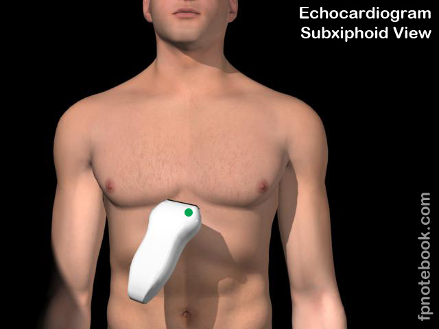
- Transducer orientation
- Hold transducer over the top (more at the base of probe) to allow for a more shallow angle to abdominal wall (15 degrees)
- Push the transducer down (posteriorly) to drop below (deep) to the xiphoid process
- Transducer placed sub-xiphoid (by 1-2 cm) in superior epigastrium
- Transducer with energy toward left Shoulder (indicator aimed at right flank)
- Regardless of screen indicator (left or right), probe indicator should match the direction of screen indicator
- Screen indicator on right (Cardiac preset, standard Echocardiogram)
- Transducer indicator pointing towards patient's left (3:00 position)
- Screen indicator on left (FAST)
- Transducer indicator pointing towards patient's right (9:00 position)
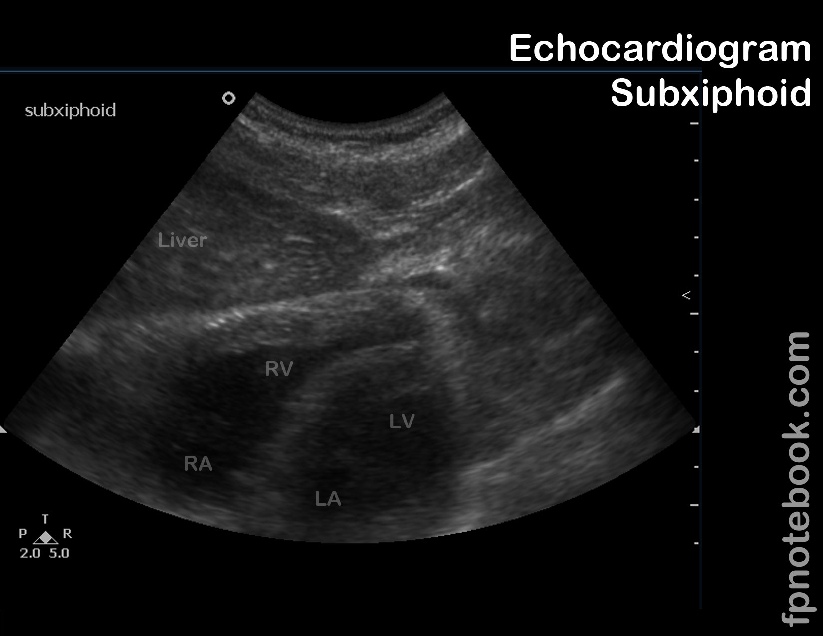
- Images
- Landmarks
- Four chamber heart view
- Increase angle of approach (aiming more posterior) if aorta is seen in the four chamber view
- Apex of the heart points to screen right when probe and screen marker are oriented correctly
- Interpretation
- Pericardial Effusion
- Systolic Dysfunction
- Wall motion abnormalities
- Resources
- FAST Exam Subcostal (Dr. Mandavia, Sonosite)
- Sub-xiphoid View Video (SonoSite)
- Echocardiographer
- References
- Palma, Bourque and Jordan (2019) Introduction to Adult Echo Ultrasound Conference, GulfCoast Ultrasound, St. Petersburg
- Mateer and Jorgensen (2012) Introduction and Advanced Emergency Medicine Ultrasound Conference, GulfCoast Ultrasound, St. Pete's Beach
- Noble (2011) Emergency and Critical CareUltrasound, Cambridge University Press, New York, p. 61-88
- Orman, Dawson and Mallin in Majoewsky (2013) EM:Rap 13(1): 4-6
- Reardon (2011) Pocket Atlas Emergency Ultrasound, McGraw Hill, New York, p. 61-106