Subtalar Dislocation, Dislocation of Subtalar Joint
- See Also
- Epidemiology
- Rare foot dislocation
- Subtalar Dislocations represent only 1 to 2% of all dislocations
- Young men account for a majority of cases
- Pathophysiology
- High energy injury (e.g. Motor Vehicle Accident, fall from height, sports such as basketball)
- Perform a full Trauma Exam on all patients
- Disruption of two joints (breaking through joint capsules and strong ligaments)
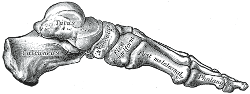
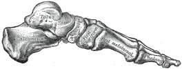 Lewis (1918) Gray's Anatomy 20th ed (in public domain at Yahoo or BartleBy)
- Talocalcaneal joint
- Talonavicular joint
- Dislocation Direction Based on Midfoot Displacement
- Medial Subtalar Dislocation (65 to 85% of cases)
- High force inversion injury while foot is plantar flexed
- Lateral Subtalar Dislocation (15 to 35% of cases)
- High force eversion injury while foot is plantar flexed
- Higher complication rate (e.g. open dislocation, interposed tissue preventing closed reduction)
- Anterior Subtalar Dislocation (rare)
- Posterior Subtalar Dislocation (rare)
- Medial Subtalar Dislocation (65 to 85% of cases)
- Signs
- Imaging
- XRay Foot
- Obtain pre-reduction and post-reduction films
- CT Foot
- Evaluate for occult associated injuries
- Associated occult injuries are common and frequently change management (e.g. ORIF)
- Fifth Metatarsal Fracture
- Talus Fracture
- Malleolus Fracture
- Osteochondral Fracture
- References
- Management
- Closed Reduction
- Perform emergently under Procedural Sedation
- Patient supine with knee flexed to 90 degrees (relaxes calf Muscles)
- Apply inline traction and countertraction
- Accentuate the deformity, and then reverse to reposition
- Apply direct pressure to talar head
- Interposed tissue may not allow for reduction (esp. lateral dislocations)
- Open reduction may be needed (one third of cases)
- Immobilization (4 to 6 weeks is typical)
- Referral
- Consult Orthopedics or podiatry for follow-up
- Emergent Consultation indications
- Open Fracture
- Neurovascular compromise
- Non-reducible dislocation
- Complications
- Open dislocation (25% of cases, esp. lateral dislocation)
- Post-Traumatic Arthritis (50-80% of cases)
- Reduced subtalar range of motion (80% of cases)
- Talus necrosis
- Subtalar Joint Stiffness
- References
- Jong and Huang (2022) Crit Dec Emerg Med 36(4): 22-3
- Lakey and Storch (2023) Crit Dec Emerg Med 37(2): 18-9
- Lugani (2022) Musculoskelet Surg 106(4):337-44 +PMID: 35435636 [PubMed]
- Prada-Cañizares (2016) Int Orthop 40(5):999-1007 +PMID: 26208589 [PubMed]