Neuron, Nerve Cell, Neural Cell, Synapse, Neuraxis Tract, Nerve Fasciculus, Neuraxis Peduncle, Neuraxis Lemniscus, Neural Nucleus, Dendrite, Neuron Cell Body, Neuron Soma, Perikaryon, Axon, Myelin Sheath, Action Potential, Nerve Impulse, Membrane Potential, Membrane Depolarization, Nerve Conduction, Nodes of Ranvier, Ranvier's Nodes, Myelinated Nerve Fiber, Saltatory Conduction, Myelination
- See Also
- Definitions
- Neuron
- Specialized conducting cell of the neurologic system which receives, conducts and transmits small electrical signals
- Building blocks of the nervous system
- Neurons are specialized into sensory Neurons and Motor Neurons
- Interneurons are a third type of Neurons that form interconnections between other Neurons
- Multiple Neurons are grouped into pathways
- Peripheral Nervous System: Nerves
- Central Nervous System: Tract, fasciculus, lemniscus, peduncle
- Synapse
- Connections between Neurons in which they communicate via chemical signals (Neurotransmitters)
- Neurologic Pathway
- Chain of communicating Neurons
- Neuraxis Tract (or fasciculus, peduncle or lemniscus)
- Bundle of axons in a pathway within the Central Nervous System (CNS)
- Nerve
- Bundle of axons in a pathway within the Peripheral Nervous System
- Neural Nucleus
- Group of Neuron cell bodies (soma) with attached group of axons (nerve tracts)
- Includes brain nucleii, Cranial Nerve nucleii, cerebellar nucleii and spinal cord nucleii
- Ganglia are the Peripheral Nerve versions of the CNS neural neuclei
- Anatomy
- Images
- Background
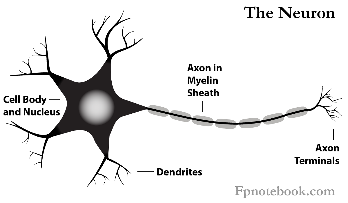
- As with all cells, Neurons have a cell body (soma) with cytoplasm and a nucleus.
- Dendrites
- Tree-like extensions along the cell body that receive signals from other Neurons, or from Sensory Receptors
- Cell Body (Soma)
- Nucleus
- Cytoplasm (Perikaryon)
- Axon
- Transmits signal from cell body to axon terminals
- From the axon terminals, the signal is passed to other nerves via Neurotransmitters across Synapses
- Myelin Sheath
- Most axons are insulated with a thin layer (myelin) of cells to conserve and speed electrical transmission
- Myelinated axons appear as white matter (while Neuron cell bodies appear as gray matter)
- Transmits signal from cell body to axon terminals
-
Neurotransmitters
- Released from axon terminals to transmit a signal into Synapse (inter-Neuron space)
- Neuron types
- Sensory Neurons
- Motor Neurons
- Interneurons (interconnections between Neurons forming a pathway)
- Group of Neurons
- Peripheral Nervous System
- Nerves
- Central Nervous System (interchangeable names)
- Nerve Tract
- Fasciculus
- Lemniscus
- Peduncle
- Peripheral Nervous System
- Physiology
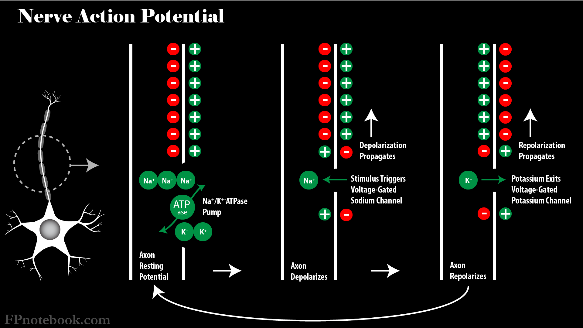
- Nerve Impulse (Action Potential)
- Neurons are specialized cells capable of tranmsitting an electrical signal
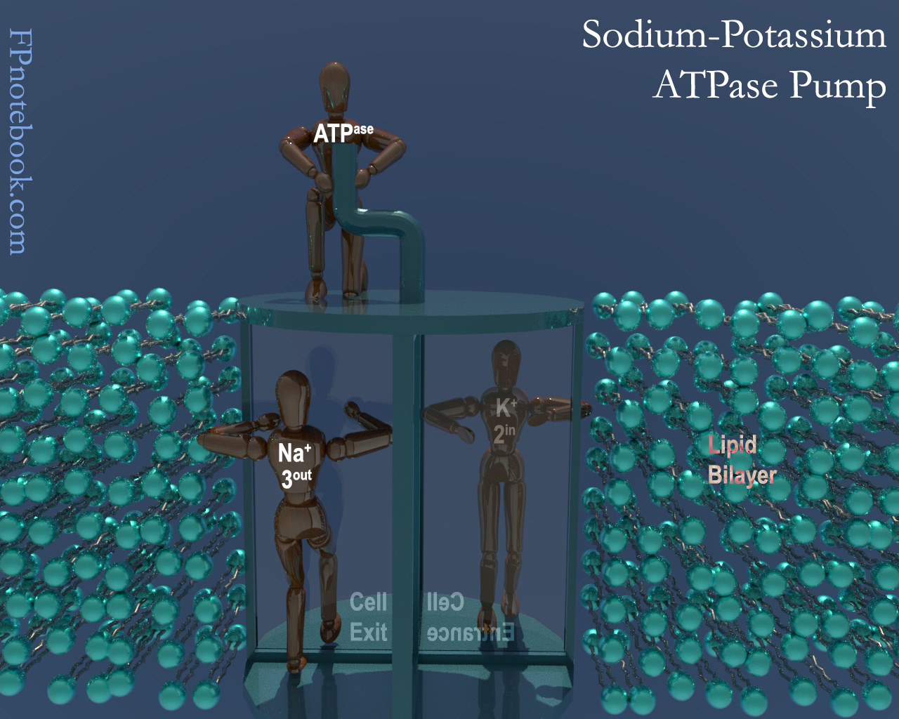
- Neuron resting Membrane Potential is more positive on outside of cell than on inside (e.g. -60 mv difference)
- Depolarization
- Voltage gated electrical channels specific for Potassium and Sodium allow for electrical signal transmission
- Voltage channels are activated when there is a neutralization of resting Membrane Potential
- Membrane Potential decreases below a threshold (e.g. 15-35 millivolts or mv)
- Nerve Depolarization is an all-or-none phenomenon
- Nerve Impulse is only initiated if there is a sufficient Action Potential
- Voltage-Gated Sodium channels suddenly open
- Sodium rushes into Neuron, resulting in neutralization of resting potential and depolarization
- Inside and outside of Neuron may have minimal difference of charge at depolarization
- Voltage-Gated Calcium channels may also be involved
- Most common in cardiac Muscle (esp. Purkinje Fibers) and Smooth Muscle (uncommon in axons)
- As with Sodium, Calcium concentrations outside the cell are higher
- When Calcium channels open, Calcium rushes into the Muscle Cell
- However Calcium channels are slower than Sodium channels
- Results in an Action Potential plateau and a delayed repolarization/recovery
- Allows for a sustained, prolonged contraction of Muscles
- Signal spreads along the axon via contiguous regions, each depolarizing in turn
- Signal amplitude is fixed regardless of the stimulus strength
- However, stronger stimuli result in increased frequency of Action Potential impulses
- Myelin Sheath
- Unmyelinated Peripheral Nerve fibers
- Axon depolarizes continuously, via contiguous ion channels along its surface
- Myelinated Peripheral Nerve fibers
- Axons are insulated, wrapped with surrounding Schwann Cells
- Gaps between the Schwann Cells are known as Nodes of Ranvier
- Ion channels are not exposed where they are covered by overlying Schwann Cells
- Ion channels are only exposed at the Nodes of Ranvier
- Action Potentials must jump between Nodes of Ranvier (Saltatory Conduction)
- Results in most faster depolarization than with unmyelinated fibers
- Axons are insulated, wrapped with surrounding Schwann Cells
- Unmyelinated Peripheral Nerve fibers
- Repolarization
- Physiology
- Synapse
- Synapse is a connection between Neurons in which they communicate via chemical signals (Neurotransmitters)
- Nerve Impulse or Action Potential (see above)
- Nerve Impulse traverses the axon until it reaches the nerve terminals
- Nerve Impulse triggers nerve terminal release of Neurotransmitters from the pre-synaptic membrane
- Neurotransmitters pass into the inter-Neuron space (Synapse)
-
Neurotransmitters
- See Neurotransmitters
- Neurotransmitters act on the post-synaptic membrane of the adjacent Neuron's Dendrites
- Neurotransmitters lower the post-synaptic Membrane Potential of the next Neuron
- Target Neuron Stimulation requires the facilitation of multiple Action Potential triggers to fire
- Stimulation of many Synapses on the same target Neuron (spatial summation) or
- Rapid succession of Action Potentials over relatively few Synapses (temporal summation)
- Each Neuron may have up to 100,000 excitatory and inhibitory inputs that, summed, determine firing potential
- Excitatory Postsynaptic potential (EPSP) refers to sum of excitatory inputs (Action Potentials)
- Inhibitory Postsynaptic potential (IPSP) refers to sum of inhibitory inputs (Action Potentials)
- Physiology
- Neuronal Networks
- Neurons are interconnected, often with thousands of inputs and outputs at a Synapse
- Neurotransmitters may have excitatory or stimulatory (positive) or inhibitory (negative) effects at the Synapse
- Patterns: Feedback Loops
- Negative Feedback
- Neuron A has excitatory effects at Neuron B
- Neuron A also has excitatory effects at Neuron C
- Neuron C inhibits Neuron A from firing
- Reverbation
- Neuron A has excitatory effects at Neuron B
- Neuron A also has excitatory effects at Neuron C
- Neuron C has excitatory effects at Neuron A, resulting in sustained firing
- Negative Feedback
- Patterns: Inter-Neuron
- Lateral Inhibition
- Neuron A, B and C lie in parallel to one another
- When Neuron B fires, it has excitatory effects on downstream Neurons
- However, Neuron B also has inhibitory effects on Neurons A and C
- Convergence
- Multiple Neurons input to a single Neuron output
- Divergence
- One Neuron has multiple Neuron outputs
- Neural Net
- Multiple Neurons interconnected with one another
- Lateral Inhibition
- Pathophysiology
- Neurotransmission Disorders
- See Neurotransmitter
- Hyperexcitable Neurons with increased automaticity
- Excessive Neurotransmitter release and activity at post-synaptic receptors
- Insufficient Neurotransmitter release and activity at post-synaptic receptors
- Images
-
 Lewis (1918) Gray's Anatomy 20th ed (in public domain at Yahoo or BartleBy)
-
 Lewis (1918) Gray's Anatomy 20th ed (in public domain at Yahoo or BartleBy)
-
 Lewis (1918) Gray's Anatomy 20th ed (in public domain at Yahoo or BartleBy)
- References
- Goldberg (2014) Clinical Physiology, MedMaster, p. 36-7, 87-9