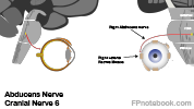
Cranial Nerve 6, Cranial Nerve VI, Abducens Nerve, CN 6, Abducens Nucleus
- See Also
- Physiology
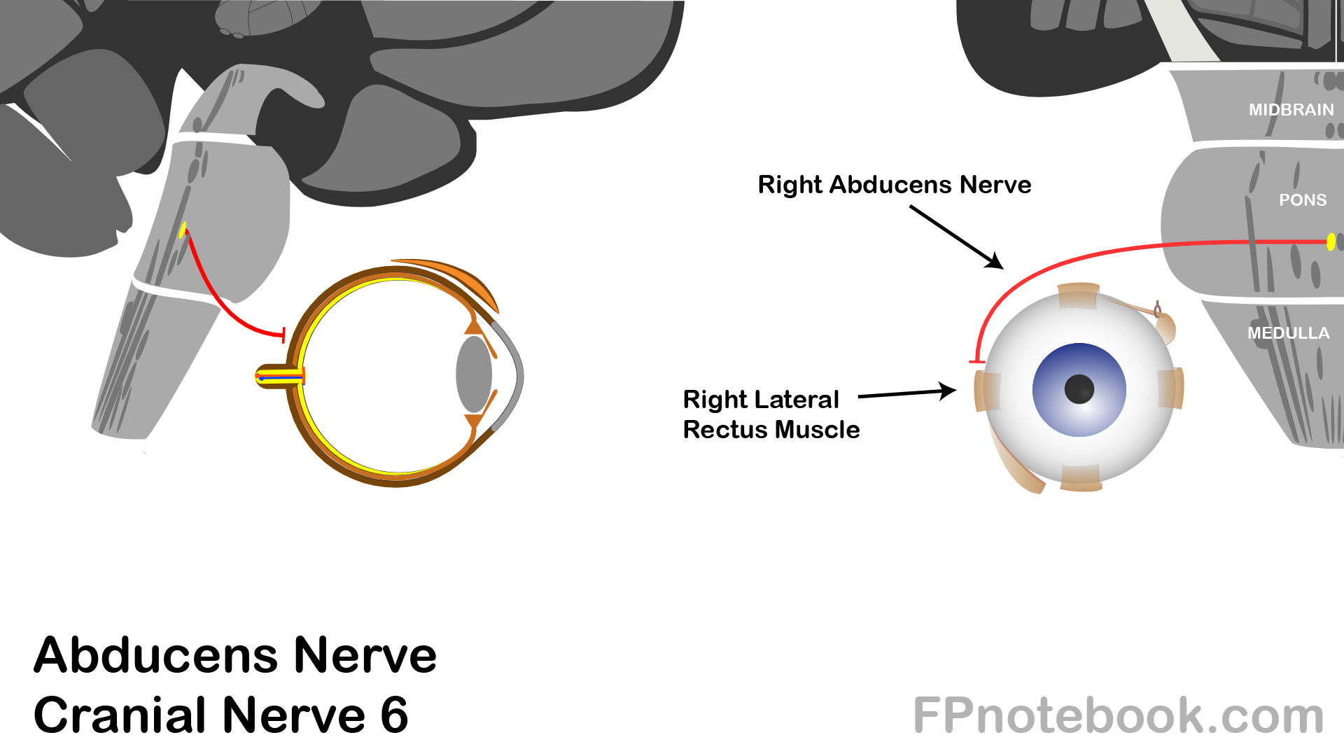
- Innervates extraocular Lateral Rectus Muscle
- Moves eye laterally
- Long thin nerve that is susceptible to compression (Cranial Nerve 4 and 6 are similar in this way)
- Paralysis (unilateral or bilateral) may occur even with generalized Increased Intracranial Pressure
- Contrast with the Cranial Nerve 3 which is a thick cable-like nerve requiring significant compression for paralysis
- Anatomy
- Abducens Nucleus
- Abducens Nucleus lies beneath facial colliculus in the pons
- Course
- As with all other Cranial Nerves (except CN 4), fibers remain ipsilateral (do not cross over)
- Nerve crosses clivus (anterior aspect of the basal portion of the Occipital Bone)
- Nerve runs below clinoid process (Sphenoid Bone)
- Nerve passes through Cavernous Sinus
- Nerve enters orbit through superior orbital fissure
- Exam
-

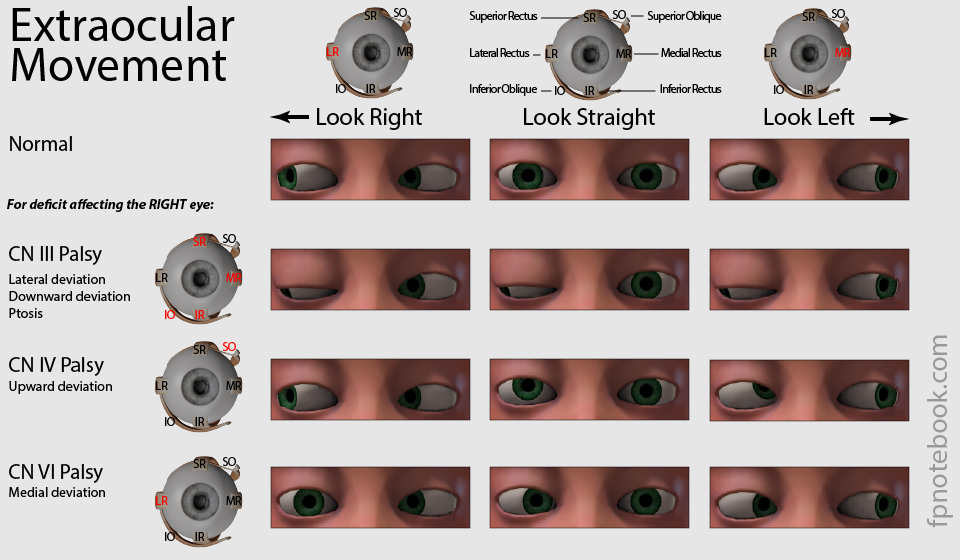
- Normal function of the Lateral Rectus Muscle
- See Extraocular Movement
- Lateral eye movement (eye abduction)
- Paralysis of the Lateral Rectus Muscle
- See Cranial Nerve 6 Palsy
- Unilateral paralysis results in lateral Gaze Palsy (may present with horizontal Diplopia)
- Causes
- CN 6 Palsy
- Distinguish from Internuclear Ophthalmoplegia (Conjugate Gaze Palsy)
-
Trauma, compression, inflammation or infection
- May occur anywhere along the CN 6 course (and nerve is thin, susceptible to injury)
- See Anatomy above
- Focal spread of infection at petrous apex of Temporal Bone (e.g. Otitis Media, Mastoiditis)
- Cavernous Sinus Thrombosis
- Cerebrovascular Accident affecting Pons
- Bilateral CN 6 Palsy Causes
- References
- Gilman (1989) Manter and Gatz Essentials of Neuroanatomy and Neurophysiology, Davis, p. 87-113
- Goldberg (2014) Clinical Neuroanatomy, p. 24-39
- Netter (1997) Atlas Human Anatomy, ICON Learning, p. 110-129