Frontal Lobe, Cerebral Frontal Lobe, Frontal Lobe Function, Primary Motor Area, Brodmann Area 4, Supplemental Motor Area, Brodmann Area 6, Frontal Eye Fields, Brodmann Area 8, Broca's Speech Area, Brodmann Area 44 and 45, Pars Orbitalis, Brodmann Area 47, Brodmann Areas of Frontal Lobe, Inferior Frontal Gyrus, Orbital Gyrus, Middle Frontal Gyrus, Superior Frontal Gyrus, Precentral Gyrus, Cingular Gyrus, Gyrus Rectus, Anterior Paracentral Gyrus
- Definitions
- Frontal Lobe
- Frontal Lobe is key to cognition, expression, problem solving and memory
- Frontal Lobe lesions may result in Expressive Aphasia, personality changes and Dementia
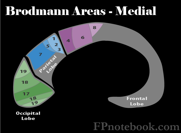
- The Primary Motor Area (Area 4), as with the Parietal Lobe's sensory regions, is organized into the cortical humunculus
- Disproportionately large region devoted to the face and hands
- Broca's Speech Area (dominant hemisphere, area 44) is key to fluent verbal expression
- Anatomy
- Brodmann Areas of Frontal Lobe
- Images
- Primary Motor Area (Area 4)
- Lesions to this area (e.g. CVA) result in Flaccid Paralysis followed by partial recovery
- Babinski Reflex may be present
- Often affected in concert with the lesions to the Supplemental Motor Area (Area 6)
- Supplemental Motor Area (Area 6)
- Lesions often affect Area 4 and Area 6 together, and result in spasticity and hyperreflexia
- Frontal Eye Fields (Area 8)
- Lesions affect voluntary Extraocular Movements looking to the opposite side
- Broca's Speech Area (Area 44, 45)
- Dominant hemisphere lesions to Broca's Speech Area results in motor Aphasia (Broca Aphasia, Expressive Aphasia)
- Patients with motor Aphasia know what they want to say, but their speech is slow and simplified
- Nouns and prepositions are often omitted
- Orbital part of Inferior Frontal Gyrus (Pars Orbitalis, Area 47)
- Involved in the processing of syntax in oral and sign languages, musical syntax, and semantic aspects of language

- Anatomy
- Gyri
- Images
 Lewis (1918) Gray's Anatomy 20th ed (in public domain at Yahoo or BartleBy)
- Precentral Gyrus
- Much of the primary motor cortex corresponds to this gyrus
- Convolution at the posterior Frontal Lobe on the convex side of both Cerebral Hemispheres
- Anterior to the Post Central Gyrus and parallel to the central sulcus
- Central sulcus separates the pre and post central gyri
- Inferior Frontal Gyrus
- Region on the surface of the Frontal Lobe visibly divided in three (pars opercularis, the pars triangularis, Pars Orbitalis)
- Bound by the inferior frontal sulcus dorsally and the lateral fissure ventrally
- Middle Frontal Gyrus
- Large region on the lateral surface of the Frontal Lobe and part of the prefrontal cortex
- Lies between the superior and the inferior frontal sulci and rostral to the Precentral Gyrus
- References
- National Cancer Institute
- Symptoms
- Findings of pathology (e.g. Tumor)
- Personality change
- Expressive Aphasia
- Signs
- Brain Lesions
- Hemiparesis (Area 4) with or without spasticity and hyperreflexia (Area 6)
- Gait disturbance
- Generalized Seizures or Focal Seizures
- Expressive Aphasia (Areas 44, 45)
- Behavior and Personality change (lesions anterior to the Primary Motor Area, Brodmann 4)
- Judgment and abstract thinking affected
- Affects Instrumental Activities of Daily Living
- May present with concerns for Dementia
- Exam
- Normal Findings (No lesions)
- Points finger each time examiner makes fist
- Makes fist when examiner points
- References
- Goldberg (2014) Clinical Physiology, Medmaster, Miami, p. 107
- Newton (1994) Am Fam Physician 49(4): 787-97 [PubMed]